ISO Metrik Face Education
Contact Us
Call Us
+1 (678) 632-3745
TESTMONIALS
ISO Metrik offers a game-changing, non-invasive solution to facial sagging — a safer, easier, and more affordable alternative to Botox and surgery. Its potential to support stroke patients with facial weakness makes it an exciting potential addition to modern stroke. rehabilitation.
Dr. Will Humphries
MAKE AN APPOINTMENT
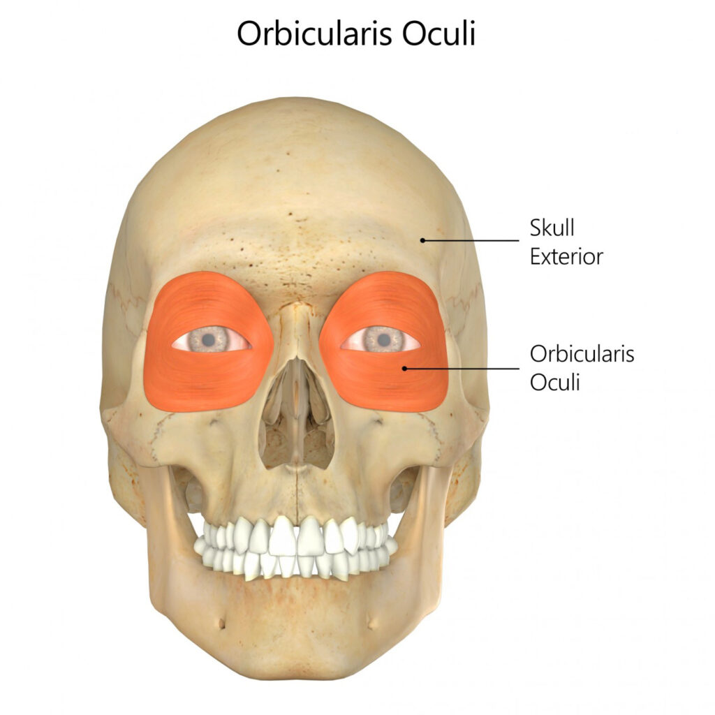
Orbicularis Oculi
Posted on 30th Jul 2020 / Published in: Face
General information
Orbicularis oculi is the muscle surrounding the eye.
Literal meaning
The little circle muscle of the eye
Interesting information
Orbicularis oculi is a skeletal muscle of the face that surrounds the eye and is responsible for closing the eye.
Injury to the orbicularis oculi can result from overuse, which may result in headaches, eyestrain, or sinus headaches. The main cause for overuse of this muscle is poor eyesight without the proper corrective lenses. If eyesight is not corrected then the eyes strain in order to try to see better, which uses the orbicularis oculi. NSAIDs, like ibuprofen, will help with headaches and proper eyewear may prevent strain. If left untreated, the orbicularis oculi can cause crow’s feet to develop.
Origin
Frontal bone.
Medial palpebral ligament.
Lacrimal bone.
Insertion
Lateral palpebral raphe.
Function
Closes the eyelids.
Nerve supply
Temporal and zygomatic branches of the facial nerve.
Blood supply
Ophthalmic artery.
Zygomatico-orbital artery.
Angular artery.
Relevant research
Previously conducted eyelid surgeries used techniques based on the premise that the nerves to the orbicularis oculi muscle approach it from the lateral and that the segmental fascicles go parallel to the muscle fibres. Surgeries based off this premise have many issues including ectropion, the flipping of the eyelids so the inward part is exposed, and other incorrect eyelid positions. An anatomical study indicated that techniques using the approach through the lower eyelid might change the innervation of orbicularis oculi muscle. This innervation change would put the muscle at a much higher risk for complications.
Ramirez, O, Santamarina, R. (2000). “Spatial orientation of motor innervation to the lower orbicularis oculi muscle”. Aesthetic Surgery Journal. 20:2, 107-113.
Blepharoptosis occurs when the upper portion of the eyelid is less than 2.0 mm from the mid-pupil or there is more than 2.0 mm of asymmetry between the two eyelids. This condition has been a challenge for surgeons due to the complications that may arise from conventional surgeries. Due to previous research, the conclusion came to develop a Frontalis-Orbicularis Oculi Muscle (FOOM) flap-shortening technique. This technique allows for the harvesting of the FOOM flaps and the adjustment of the flaps depending on the severity of the condition. The procedure prevents the eye from closing or drooping involuntarily and enhances the ability to keep the eye wide open. This study showed that the procedure yielded good results with the biggest issue being under-correction.
Lai, C, Lai, C, Huang, S, Feng Sun, I, Chang, K, Lee, S, Lin, S. (2010). “A new trend for the treatment of blepharoptosis: Frontalis-Orbicularis Oculi Muscle flap shortening technique”. Journal of Plastic, Reconstructive & Aesthetic Surgery. 63:2, 233-239.
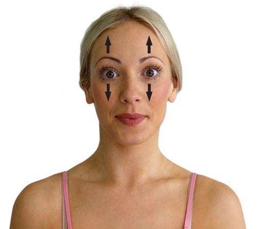
Orbicularis oculi exercises
The eyelid squeeze
Begin by placing the fingertips on the eyebrows. Close the eyelids and squeeze tightly. Hold the position for forty seconds. Practice this exercise five times a day.
Surprise eyes
Begin sitting with good posture. Raise the eyebrows up as far as possible and open the eyes widely. Hold this position for ten seconds, release for five seconds, and then repeat ten times. Perform this exercise daily.
The Orbicularis Oculi: Strength Begins at the Maxilla–Zygomatic Connector
The orbicularis oculi muscle encircles the eye and is essential for blinking, squinting, and expressive communication. While the muscle itself wraps around the eye socket, its form and function depend on the strength of the bones beneath it—most notably, the maxilla (upper jaw) and zygomatic bone (cheekbone).
But it’s not just the individual bones—it’s the connector between them that determines the stability, tone, and contour of the entire midface and orbital region.
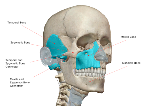
The Critical Role of the Maxilla–Zygomatic Connector
Where the zygomatic bone meets the maxilla, a structural bridge is formed. This connector:
- Serves as the foundation for muscles like the orbicularis oculi, zygomaticus major, and zygomaticus minor
- Supports the lower eyelid and cheek fat pads
- Maintains the projection and volume of the under-eye region
- Preserves the natural frame and lift around the eye
When this bony junction begins to degrade—whether from aging, trauma, or lack of muscular engagement—muscles in the region begin to sag or atrophy. This results in:
- Under-eye hollowing
- Crow’s feet and fine lines
- Drooping of the lower eyelid
- Flattened or sunken cheek contours
- A less defined, aged midface appearance
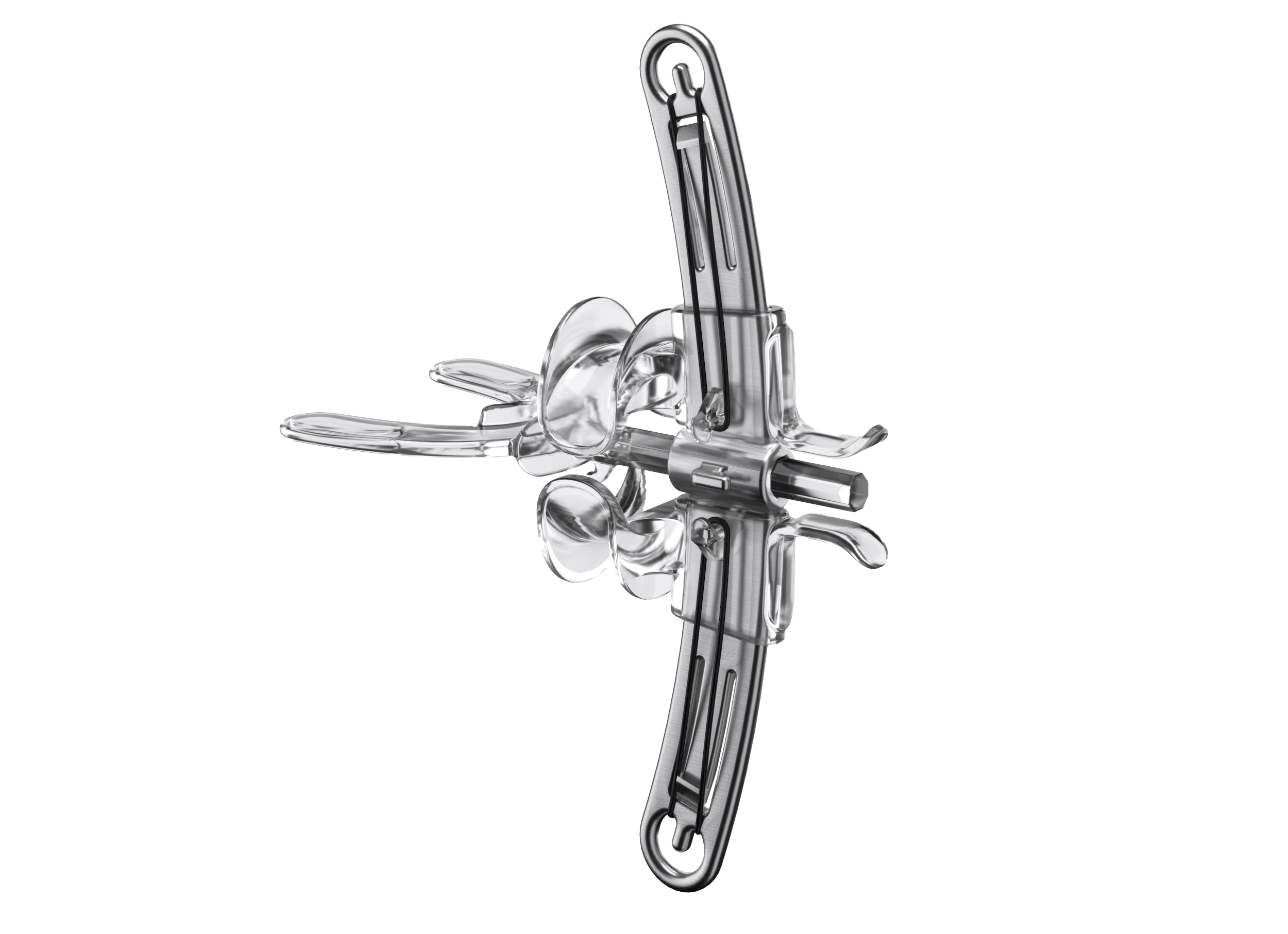
The ISO Metrik Device: Activating the Muscles That Reinforce the Connector
The ISO Metrik Device was engineered to indirectly reinforce the maxilla–zygomatic connector by training the muscles that originate or insert near this junction—specifically the zygomaticus major and minor.
Here’s how the ISO Metrik Device plays a key role:
- Applies internal resistance that stimulates the zygomatic system, strengthening the muscular tension that supports the midface structure
- Promotes cheek lift, which naturally reduces pull on the lower eyelid
- Improves upper lip and smile elevation, enhancing harmony with the orbicularis oculi
- Helps restore tension at the connector, reducing midface collapse and redefining the under-eye region
Why the Connector Matters
The maxilla–zygomatic junction isn’t just a point of contact—it’s the keystone of facial architecture in the midface. When it weakens, the entire facial system loses cohesion. When it’s supported—through muscular activation and resistance—it reestablishes the natural lift, contour, and expression of the face.
The ISO Metrik Device offers a non-invasive way to rebuild and reinforce this critical junction, helping you reclaim the youthful tone and balance that begins with your eyes.
Want to refresh your eyes and lift your midface—naturally and from within?
Start at the source: the maxilla–zygomatic connector, activated through the ISO Metrik Device.
TESTMONIALS
Dimply dummy text of the deaprinting and typesetting industryorem Ipsum has been the industry’s standard dummy dearty.
DAVID SMITH
CEO,Radiustheme
Contact Us
Call Us
+1 (678) 632-3745
MAKE AN APPOINTMENT
Your journey to facial rejuvenation begins here. Join the ISO Metrik Device Community today.